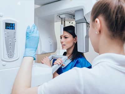
Digital radiography replaces traditional film with electronic sensors and computer systems to capture, display, and store dental X-ray images. Instead of chemically developed film, a small sensor or phosphor plate records the image and transmits it to a computer where it becomes immediately viewable. This shift from analog to digital has transformed routine imaging from a time-consuming task into a seamless part of modern dental care.
The technology is not a single device but a set of tools that work together: intraoral sensors for bitewing and periapical images, software that processes and enhances pictures, and secure storage systems that keep records accessible. Because images are created and managed digitally, clinicians can integrate them with electronic health records, treatment-planning software, and other diagnostic tools to form a more complete picture of oral health.
For patients, the most noticeable differences are speed and clarity. Images appear on-screen within seconds, which accelerates clinical decisions and improves communication between the dental team and the person in the chair. While the core purpose of radiography—revealing structures not visible during an exam—remains the same, digital methods make those details easier to see and act on.
One of the primary advantages of digital radiography is image quality. Modern sensors capture fine detail and software tools allow clinicians to adjust contrast, zoom in on areas of interest, and measure distances with precision. These enhancements make it easier to detect small cavities, subtle bone changes, and the early signs of conditions that might otherwise be missed on conventional film.
Because the images are available instantly, dentists can compare current and prior images side-by-side without waiting for film processing. This temporal comparison is valuable for monitoring progression or healing over time. The ability to annotate, magnify, and manipulate images supports clearer treatment explanations and more confident diagnoses.
Digital systems also reduce the likelihood of retakes. When an image needs adjustment, clinicians can fine-tune settings or reposition the sensor and capture a new image quickly. Fewer retakes mean less inconvenience and a smoother appointment for patients, while the team benefits from a more efficient workflow and better-quality clinical records.
Advances in sensor sensitivity and image processing mean that digital radiography generally requires less radiation than older film-based methods. While any use of ionizing radiation is carefully controlled, modern digital systems enable clinicians to obtain diagnostic-quality images with reduced exposure whenever appropriate. This improvement aligns with the widely accepted principle of using the lowest radiation necessary to achieve clear diagnostic results.
Beyond reduced radiation, digital imaging contributes to safety in other practical ways. Because images are captured and displayed instantly, procedures tend to be faster, and patient time in the operatory is minimized. Digital records are also stored electronically, limiting the need to handle physical films and reducing the risk of damage or loss.
From a staff perspective, handling digital images avoids exposure to developing chemicals and other materials used in film processing. The elimination of chemical processing also has environmental benefits, removing the need to dispose of processing fluids and paper materials associated with traditional radiography.
Digital radiography plays a central role in modern treatment planning. Clear, manipulable images integrate directly with charting systems, 3D imaging tools, and restorative planning software to guide procedures ranging from routine fillings to complex implant placement. Clinicians can take measurements, assess root anatomy, and evaluate bone margins with tools built into digital imaging software.
Because digital images are easy to share, coordinating care with specialists is more straightforward. Images can be exported or securely transmitted to an endodontist, oral surgeon, or orthodontist for consultation without the delay associated with mailing films or making physical copies. This connectivity supports timely referrals and a more coordinated treatment approach.
Digital images also enhance patient education. Displaying radiographs on a monitor during the visit allows the dental team to point out areas of concern, explain treatment options, and show anticipated outcomes. When patients see the same images their dentist is using to plan care, they are better informed and more engaged in decisions about their oral health.
Moreover, predictable, high-quality imaging contributes to clinical documentation. Well-organized digital records support continuity of care, whether a patient returns to the same office or seeks treatment elsewhere. Clear imaging documentation is an important part of evidence-based treatment planning and follow-up.
In a typical dental appointment, digital radiography is used to augment the visual exam and reveal structures hidden from plain sight. Bitewing images help detect interproximal cavities and evaluate bone levels, while periapical images show root structure and the tips of the tooth roots. These views are commonly used during routine checkups, restorative planning, and assessment of dental pain or infection.
Clinicians also rely on digital images for pre-treatment assessments before restorative work or surgical procedures. For example, careful radiographic evaluation helps confirm root canal anatomy, assess the footprint of a failing restoration, or evaluate the bone available for an implant. Because images can be adjusted and measured within the software, planning is both more precise and more predictable.
Within the practice, digital radiography supports efficient recordkeeping and communication. Images are stored securely in the patient’s electronic file and can be revisited during later visits or included with referral documentation. The convenience of digital files simplifies appointment workflows and preserves a consistent visual history of oral health over time.
In summary, digital radiography is a cornerstone of modern dental care—delivering clearer images, greater safety, and faster, better-informed clinical decisions. At Smiles Dental at Reston Town Center, we incorporate digital imaging into our diagnostic and treatment processes to improve patient comfort and outcomes. If you have questions about how digital X-rays are used in your care, please contact us for more information.


Digital radiography uses electronic sensors or phosphor plates and computer software to capture dental radiographs instead of traditional film. Images appear on a monitor within seconds, allowing clinicians to review and adjust images during the appointment. This change has streamlined workflow, improved diagnostic clarity, and reduced the time patients spend waiting for results.
Because images are digital, they can be enhanced, measured, and compared with prior studies to support more precise treatment decisions. Digital files integrate with electronic health records and treatment-planning tools, enabling a more comprehensive view of oral health. At Smiles Dental at Reston Town Center, we incorporate digital imaging into routine diagnostics to improve patient comfort and clinical outcomes.
Digital radiography typically employs intraoral sensors for bitewing and periapical views and plate-based systems for similar intraoral images. Bitewings are used to evaluate interproximal tooth surfaces and crestal bone, while periapical views reveal root structure and the tips of the tooth roots. Some practices also use extraoral digital sensors for panoramic views, depending on the diagnostic need.
Sensors vary in size and flexibility to accommodate different mouths and clinical situations, and phosphor plate systems offer a thinner alternative that can be scanned after exposure. The choice of sensor and view depends on the clinical question, patient comfort, and the level of detail required for treatment planning. Clinicians select the appropriate combination to obtain diagnostically useful images while minimizing patient discomfort.
Digital systems produce high-resolution images that clinicians can enhance by adjusting contrast, brightness, and magnification to reveal subtle details. Software tools also allow precise measurements of distances and angles, which are helpful for implant planning, root canal assessment, and evaluating bone levels. These capabilities help identify early-stage cavities, small fractures, and minor bone changes that might be missed on conventional film.
Instant comparison of current and prior images supports monitoring of healing, disease progression, or the success of a restoration over time. Annotating and saving enhanced views in the patient record improves communication among members of the dental team and with outside specialists. Overall, digital radiography contributes to more predictable, evidence-based treatment decisions.
Advances in sensor sensitivity and image processing allow diagnostic-quality radiographs at lower exposure levels than older film systems. While any use of ionizing radiation is performed only when clinically indicated, digital radiography enables clinicians to follow the principle of ALARA—keeping exposure as low as reasonably achievable. Reduced exposure does not compromise image quality when appropriate techniques and settings are used.
Digital imaging also eliminates the need for chemical processing, removing staff contact with developers and disposables associated with film. Faster image acquisition shortens the time patients spend in the operatory, which can reduce overall procedural time and potential discomfort. Together, these improvements offer practical safety and environmental benefits in addition to lower radiation doses.
Digital radiographs are saved directly into a secure electronic record and organized with the patient file for easy retrieval during future visits. Images can be exported in standard formats or transmitted securely to specialists, laboratories, or referring clinicians for timely consultation. This avoids the delays and physical handling associated with transporting film or paper copies.
When sharing images, practices use encrypted channels or secure portals to protect patient information and comply with privacy regulations. Digital storage also supports versioning and long-term archival so clinicians can review a consistent visual history of a patient’s oral health. Efficient sharing and storage promote coordinated care and faster treatment planning when multidisciplinary input is needed.
Yes. Immediate image preview allows clinicians to assess image quality right away and make adjustments while the patient is still positioned, reducing the likelihood of repeat exposures. Software enhancements can correct minor contrast or brightness issues without requiring a new radiograph, which further decreases the number of retakes. When a retake is necessary, adjustments can be made quickly to avoid repeated patient visits.
Sensor technology and improved positioning aids also contribute to fewer repeats by producing more consistent, high-quality images on the first try. Fewer retakes benefit patients by minimizing cumulative exposure and shortening appointment times. For the clinical team, reduced retakes improve efficiency and maintain better-quality records.
Patients typically notice faster appointments because images are captured and available immediately, eliminating film processing delays. The ability to view radiographs on a monitor helps clinicians explain findings visually, which can increase patient understanding and engagement in care. Smaller, more comfortable sensors and fewer repeat exposures can make imaging less intrusive for many patients.
Because images are digital, clinicians can enlarge areas of concern and annotate them during the visit to show why specific treatments are recommended. This visual approach supports clearer conversations about diagnosis and options without relying solely on technical descriptions. Overall, digital radiography often makes imaging quicker, clearer, and more informative for patients.
Absolutely. Digital files are ideal for longitudinal comparison because current images can be displayed side-by-side with prior studies to assess changes in tooth structure, restorations, or bone levels. This temporal perspective is valuable for tracking the progression of cavities, periodontal disease, healing after treatment, or the stability of restorations and implants. Consistent imaging protocols allow clinicians to make accurate comparisons across visits.
Measurements and annotations saved in the record provide objective documentation that supports evidence-based follow-up and decision-making. Regular radiographic monitoring is used selectively based on individual risk factors, clinical findings, and established guidelines. When changes are detected early, clinicians can intervene sooner and often with simpler treatments.
Digital radiography generally refers to two-dimensional intraoral and extraoral radiographs that are excellent for routine diagnosis, cavity detection, and basic treatment planning. Cone beam computed tomography, or CBCT, provides three-dimensional imaging that is useful for complex surgical planning, implant placement in difficult anatomy, and assessment of certain pathologies. Each modality has strengths and is selected based on the diagnostic question at hand.
In practice, clinicians often use digital radiographs for initial evaluation and reserve CBCT for cases that require detailed 3D visualization. Digital systems integrate so 2D and 3D images can be reviewed together in treatment-planning software when appropriate. This complementary approach allows clinicians to choose the most informative, least invasive imaging for each patient.
Digital radiographs are stored in the dental practice’s secure electronic record system with access controls that limit who can view patient files. Standard safeguards include encrypted data storage, user authentication, and routine backups to preserve records while preventing unauthorized access. These measures help ensure that radiographic images are treated as protected health information under applicable privacy regulations.
When images are shared with outside providers, transmission uses encrypted methods or secure portals to maintain confidentiality during transfer. Staff training and established protocols govern how images are handled, retained, and disposed of to maintain compliance and protect patient privacy. Our office, Smiles Dental at Reston Town Center, follows these practices to safeguard your imaging records.

Ready to schedule your next dental appointment or have questions about our services?
Contacting Smiles Dental at Reston Town Center is easy! Our friendly staff is available to assist you with scheduling appointments, answering inquiries about treatment options, and addressing any concerns you may have. Whether you prefer to give us a call, send us an email, or fill out our convenient online contact form, we're here to help. Don't wait to take the first step towards achieving the smile of your dreams – reach out to us today and discover the difference personalized dental care can make.