Digital impressions have transformed how dentists capture the shape and detail of teeth and surrounding tissues. At Smiles Dental at Reston Town Center, we use advanced intraoral scanners to replace traditional putty-based molds with fast, precise digital records. The result is a smoother, more comfortable experience for patients and a highly accurate foundation for restorative and cosmetic dentistry.
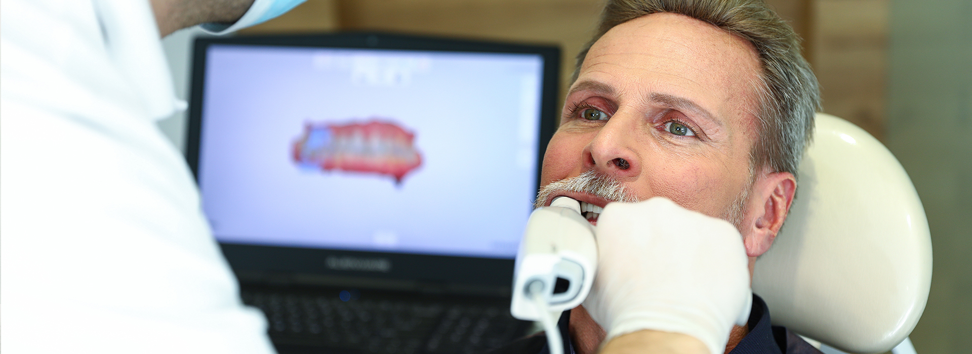
Intraoral scanners are handheld devices that record hundreds to thousands of images per second as they pass over teeth and gums. Specialized software stitches those images together to build a three-dimensional model that represents the patient’s mouth in fine detail. This process bypasses the need for physical impression trays and the impression materials used in conventional techniques.
The scanner captures surface textures, margins, and interproximal contacts with remarkable fidelity, enabling clinicians to evaluate anatomy on-screen and make immediate adjustments. Because the data is digital from the start, it can be annotated, measured, and integrated with other diagnostic tools such as digital X-rays and CBCT scans.
Clinically, the digital scan becomes the starting point for planning restorations, orthodontic alignments, and implant placements. The technology emphasizes repeatable accuracy: scans can be reviewed in real time, and additional passes can be taken immediately if any area needs clarification instead of rescheduling the patient for a remake.
One of the most noticeable advantages of digital impressions is the improved comfort for patients. Traditional impressions often require bulky trays and impression material that can trigger gagging, taste issues, or simply prolonged discomfort. Digital scans eliminate those inconveniences by capturing the required information with a quick, noninvasive wand.
For patients with sensitive gag reflexes, limited mouth opening, or anxiety about dental procedures, intraoral scanning is often perceived as less intimidating. The scanning process is usually shorter than traditional impression appointments, and team members can pause at any time to check the patient’s comfort or reposition the scanner.
Beyond immediate comfort, patients benefit from clearer communication: clinicians can show the scan on a monitor, point out areas of concern, and walk patients through proposed treatments using the same visual data. This transparency helps patients understand their diagnosis and treatment options without relying on abstract descriptions.
Digital impressions provide a level of detail that supports precise restorative work. By accurately capturing margins, occlusal relationships, and adjacent tissues, scans reduce the risk of ill-fitting crowns, bridges, or inlays. This precision helps labs and CAD/CAM systems fabricate restorations that seat properly and require fewer adjustments at try-in.
Because digital models can be measured and reviewed before manufacturing, clinicians can identify potential issues early—such as insufficient clearance or a challenging margin—and address them in the same appointment. That proactive approach minimizes remakes and streamlines the path from preparation to final restoration.
When integrated with design software, digital impressions enable customized restorations tailored to a patient’s anatomy and bite. Whether the final restoration is crafted in an outside dental laboratory or manufactured in-office with CAD/CAM milling, starting from a high-quality digital file improves predictability and long-term fit.
Digital files travel instantly to dental laboratories or in-office milling systems, removing delays associated with shipping physical impressions and pouring stone models. Electronic transmission of high-resolution scans reduces transit time and the chance of damage or distortion that can occur with physical impressions.
Laboratories that receive digital scans can begin designing restorations sooner and often with greater confidence, since they’re working from consistent, measurable data. Digital workflows also support version control: teams can save and resend files without losing fidelity, and records remain accessible for future reference or modifications.
For the dental team, this efficiency translates into better scheduling and fewer follow-up visits for impression-related remakes. The streamlined exchange between clinic and lab encourages closer collaboration on complex cases and supports contemporary treatment timelines that many patients prefer.
Digital impressions are a cornerstone of same-day restorative systems. When combined with in-office CAD/CAM milling, a high-quality scan can be used immediately to design and fabricate ceramic restorations during a single appointment. This capability reduces the number of visits required to complete crowns, onlays, or veneers while maintaining laboratory-grade materials and aesthetics.
Integration with other digital technologies—such as digital radiography and 3D imaging—creates a comprehensive picture of oral health. This ecosystem allows dentists to plan restorations with an awareness of underlying bone structure, root positions, and occlusal dynamics, making treatments more predictable and conservative when possible.
Beyond restorations, digital impressions aid in interdisciplinary care. Clear files can be shared with orthodontists, periodontists, and implant specialists to coordinate treatment planning, ensuring that every provider works from the same accurate dataset.
What patients can expect during and after a scan
Preparing for a digital scan usually requires minimal to no special instructions. During the appointment, the clinician will gently move the scanner around the teeth and soft tissues; patients may be asked to reposition their tongue or slightly open their mouth to assist with access. The process is quick, typically completed within a few minutes for a single arch depending on the complexity of the case.
After the scan, the dentist will review the model on-screen, explain any findings, and discuss the recommended next steps. If a restoration is being planned, the digital data will be sent electronically to the lab or to an in-office milling system. Patients should expect follow-up based on the chosen workflow—either return for restoration placement or remain for same-day fabrication when available.
Because the scan is noninvasive and requires no setting materials, there’s no physical aftercare specifically related to the impression. Routine precautions related to the preparatory procedure—such as temporary crown care or soft-tissue healing—will be covered by the clinician as appropriate.
In summary, digital impressions represent a meaningful improvement in accuracy, comfort, and efficiency for modern dental care. At Smiles Dental at Reston Town Center, we leverage this technology to provide precise diagnostics and to streamline restorative workflows while focusing on patient comfort. If you’d like to learn more about how digital scanning could apply to your treatment, please contact us for more information.

At Smiles Dental at Reston Town Center, digital impressions are high-resolution three-dimensional records of the teeth and soft tissues captured with an intraoral scanner rather than putty-based impression materials. The scanner records hundreds to thousands of images per second and software stitches them into a precise 3D model that represents the patient’s mouth in detail. Unlike traditional trays and impression compound, the digital file is immediately available for review, measurement, and electronic transfer to a laboratory or milling unit.
Digital impressions serve as the starting point for restorations, orthodontic aligners, and implant planning because they capture margins, occlusal relationships, and adjacent anatomy with consistent fidelity. The digital workflow reduces the risk of physical distortion that can occur when impressions are shipped, poured, or handled. Clinicians can annotate scans, make adjustments on-screen, and archive files for future reference without producing stone models unless specifically required.
An intraoral scanner is a handheld wand that the clinician passes over the teeth and gums while specialized software captures sequential images or point clouds and constructs a continuous 3D model. The clinician typically scans quadrants or entire arches in a single visit, reviewing the reconstruction in real time to ensure complete coverage and clear margins. If an area needs refinement, additional passes are taken immediately so the scan is complete before the patient leaves the operatory.
The digital data can be measured and analyzed on-screen, allowing clinicians to check occlusal clearance, identify undercuts, and evaluate preparation margins before sending files to a lab or CAD/CAM system. Scans are inherently compatible with other digital tools, so they can be combined with intraoral photos, digital X-rays, and CBCT datasets for comprehensive treatment planning. This immediate feedback shortens the workflow and reduces the likelihood of remakes caused by incomplete or distorted impressions.
Digital impressions eliminate the need for bulky trays and setting materials that can cause gagging, unpleasant tastes, or anxiety for many patients. The scanner is a small, noninvasive wand that captures images quickly, and team members can pause or reposition at any time to improve patient comfort. For patients with limited mouth opening, sensitive gag reflexes, or dental anxiety, scanning is often perceived as less intrusive and faster than conventional impressions.
Beyond physical comfort, digital scans improve communication by allowing clinicians to show patients a visual model of their teeth and soft tissues on a monitor. This visual information helps patients understand diagnoses and treatment options more clearly than abstract descriptions alone. The transparency created by on-screen reviews often leads to better-informed decisions and greater patient confidence in the proposed plan.
Digital impressions capture fine details such as preparation margins, interproximal contacts, and occlusal surfaces with high repeatability, which supports better-fitting crowns, bridges, inlays, and onlays. Because clinicians can measure and inspect the model before manufacturing, they can identify potential issues like insufficient reduction or challenging margins and address them immediately. That preemptive review reduces the frequency of ill-fitting restorations and the need for multiple try-ins.
When used with CAD/CAM design software, high-quality scans enable customized restorations that match a patient’s anatomy and occlusion, improving long-term fit and function. Whether a restoration is milled in-office or fabricated by a dental laboratory, starting from an accurate digital file enhances predictability and minimizes chairside adjustments. This precision supports efficient restorative workflows and helps maintain occlusal harmony and marginal integrity over time.
Yes. When combined with in-office CAD/CAM design and milling systems, a high-quality digital scan can be used to design, mill, and place ceramic restorations within a single appointment. The digital workflow allows the clinician to capture the preparation, design the restoration with integrated software tools, and fabricate the final piece in porcelain or other restorative materials without relying on external lab shipping. This approach reduces the number of patient visits while delivering laboratory-grade materials and aesthetics.
Not every case is suitable for same-day fabrication; complex multi-unit restorations, extensive occlusal rehabilitation, or certain material preferences may still require laboratory collaboration. In those situations the digital file is transmitted electronically to a trusted dental lab to continue the workflow, which still benefits from the speed and accuracy of the original scan. The clinician determines the best pathway based on case complexity and clinical objectives.
Digital scans are exported as standardized file formats and sent electronically to dental laboratories or in-office milling systems, eliminating the delays and potential distortions associated with shipping physical impressions. Electronic transmission is rapid and preserves the file’s resolution, which allows laboratory technicians to begin design work sooner and with greater confidence in the digital dataset. Many labs support secure upload portals or direct integration with office software to simplify the exchange process.
Because digital workflows maintain version control, teams can save, revisit, and resend files without degradation, which aids collaboration on complex cases and supports efficient revisions when needed. The digital archive also streamlines communication between specialists—such as orthodontists, periodontists, and implant surgeons—who can work from the same accurate dataset. This interoperability improves coordination and reduces the chance of misinterpretation during restorative or interdisciplinary treatment planning.
Although digital impressions are highly versatile, there are clinical situations where conventional impressions remain useful, such as when extensive soft-tissue bleeding, heavy saliva, or certain subgingival margin conditions limit scanning visibility. Some laboratories or specific restorative protocols may request physical models for particular workflows or material processes that currently depend on stone models. In pediatric cases or very uncooperative patients a clinician may elect a conventional technique when it provides a more predictable result for that appointment.
Clinicians often use a hybrid approach, combining digital scans with selective traditional techniques when appropriate to capture hard-to-access anatomy or to meet laboratory requirements. The choice between digital and analog methods is guided by clinical judgment, case complexity, and the desired restorative outcome. The priority is always to select the impression method that ensures accurate records and a reliable final restoration.
Preparation for a digital scan is minimal; patients usually need no special instructions beyond routine oral hygiene and any directions related to a specific restorative procedure. During the appointment the clinician will pass the scanner gently over the teeth and surrounding tissues, and the process typically takes only a few minutes per arch depending on case complexity. Patients may be asked to reposition their tongue or open wider briefly to improve access, and team members will pause as needed to maintain comfort.
After the scan the dentist will review the 3D model on-screen, discuss findings, and explain next steps, whether that means sending the digital data to a laboratory or proceeding with in-office fabrication. There is no physical aftercare related to the scanning itself, though any instructions tied to the preparatory treatment—such as temporary crown care or soft-tissue healing—will be provided. The practice will schedule follow-up visits according to the chosen workflow and expected fabrication timeline.
Digital impressions can be superimposed on CBCT datasets and digital radiographs to create a comprehensive three-dimensional treatment plan that includes soft-tissue morphology and underlying bone anatomy. This fusion of surface scans with volumetric imaging is especially valuable for implant planning, where accurate prosthetic-driven placement improves long-term outcomes. Integration also supports restorative and orthodontic workflows by providing a complete view of occlusion, root positions, and bone relationships.
Specialized software aligns the different datasets so clinicians can simulate restorations, evaluate implant angulation, and coordinate interdisciplinary care with greater precision. Sharing these integrated files with referring specialists ensures everyone works from the same visual and measurable information. The result is more predictable planning, conservative surgical execution, and restorations that respect both function and esthetics.
Digital scans are stored as part of the patient’s electronic record using secure systems that support encrypted transfer and controlled access to preserve privacy and data integrity. Offices generally use accepted clinical record systems or laboratory portals that track file versions, user access, and modifications so scans remain auditable and retrievable for future treatment. Secure storage also simplifies long-term care by keeping baseline anatomy on file for comparisons, repairs, or future restorative work.
At Smiles Dental at Reston Town Center we maintain digital records to support continuity of care and interdisciplinary collaboration while limiting access to authorized clinical staff. Patients can expect their scans to be available for follow-up treatments, specialist referrals, and future restorations without the need for repeated impressions unless clinical changes warrant an updated scan. The practice follows established protocols for data handling to ensure records remain accurate, secure, and useful for ongoing care.

Ready to schedule your next dental appointment or have questions about our services?
Contacting Smiles Dental at Reston Town Center is easy! Our friendly staff is available to assist you with scheduling appointments, answering inquiries about treatment options, and addressing any concerns you may have. Whether you prefer to give us a call, send us an email, or fill out our convenient online contact form, we're here to help. Don't wait to take the first step towards achieving the smile of your dreams – reach out to us today and discover the difference personalized dental care can make.