Advanced imaging plays a central role in modern dentistry, and cone-beam computed tomography (CBCT) has reshaped how clinicians evaluate anatomy, plan procedures, and communicate with patients. CBCT captures three-dimensional views of the teeth, jaws, and surrounding structures in a single scan, revealing details that conventional two-dimensional X-rays can miss. This richer perspective helps clinicians identify issues earlier and design treatments with greater precision.
At Smiles Dental at Reston Town Center, our team incorporates CBCT into cases where three-dimensional information adds clear clinical value. We apply the technology selectively—when the diagnostic benefit outweighs any additional exposure—and pair the scans with a thorough clinical exam to form a complete picture of each patient’s oral health. The result is a more confident diagnosis and a treatment plan tailored to the individual.
Below are practical, patient-friendly explanations of what CBCT does, how we use it, and what you can expect when a scan is recommended. The goal is to demystify the technology while demonstrating how it supports safer, more predictable dental care.
CBCT produces volumetric images that show structures in three dimensions, giving clinicians the ability to view teeth, roots, bone, and adjacent anatomy from multiple angles. This level of detail makes it easier to detect root fractures, evaluate complex tooth anatomy, and locate impacted teeth. For conditions that are ambiguous on traditional X-rays, a CBCT scan often provides the decisive information needed to move forward confidently.
Because CBCT data can be reformatted into cross-sectional slices, clinicians can precisely assess the thickness and density of bone in specific regions. That’s especially valuable when considering treatments that interact closely with bone or neural structures, such as implant placement or surgical extractions. The images also help identify anatomical variations—like sinus proximity or nerve pathways—that could influence treatment decisions.
Importantly, the diagnostic benefit of CBCT extends beyond individual teeth. The scan can reveal hidden pathology, asymmetries, or developmental issues in the jaws and facial bones that might not be evident on 2D films. When used judiciously, CBCT reduces uncertainty and supports more informed, conservative care planning.
CBCT has a broad range of dental applications. In implant dentistry, three-dimensional imaging is instrumental for mapping available bone, determining ideal implant size and angulation, and avoiding important anatomical structures. For endodontic concerns, CBCT can reveal complex root canal systems, missed canals, or the extent of periapical lesions, guiding targeted treatment rather than guesswork.
Oral surgery and impacted-tooth management benefit from CBCT’s spatial accuracy, which helps surgeons plan incisions, access points, and the safest path for tooth removal. Orthodontists also use CBCT to evaluate jaw relationships, airway volume, and tooth position when standard records are insufficient. Additionally, CBCT can aid in the assessment of temporomandibular joint (TMJ) conditions and in detecting certain benign or pathological lesions that require follow-up or referral.
Because each application has distinct imaging needs, the scan volume and resolution can be adjusted to optimize visualization for the clinical question at hand. Tailoring the scan reduces unnecessary exposure while maximizing the diagnostic return for the specific procedure under consideration.
Radiation safety is an important consideration for any imaging modality. Modern CBCT units include low-dose protocols and the ability to limit the scanned field of view to the smallest region necessary, which helps minimize exposure. Our team follows guiding principles—such as only recommending scans when they will influence care—and uses settings appropriate for the diagnostic task.
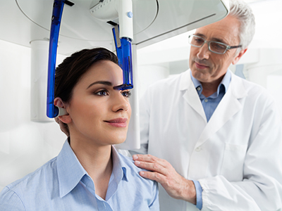
The scanning process itself is quick and noninvasive. Most CBCT examinations take under a minute of active imaging time, and patients remain seated or standing in an open design that is generally well tolerated. There is no intravenous contrast, no confinement in a tunnel as with some medical CT scanners, and no recovery time required. After the scan, the dataset is available immediately for review and planning.
We also prioritize clear communication about why a scan is recommended and what the images will be used for. Understanding the purpose and the steps involved helps patients feel more comfortable and engaged in their care decisions.
One of the strengths of CBCT is how easily its digital datasets integrate with planning software and other imaging modalities. In implant therapy, for example, CBCT scans can be combined with digital impressions to design surgical guides that translate virtual plans into predictable, guided placement. This digital workflow improves surgical precision and can shorten chair time during implant placement.
CBCT files are also valuable when coordinating care with specialists. A detailed 3D dataset can be shared with oral surgeons, periodontists, endodontists, or orthodontists, enabling collaborative planning without redundant imaging. When pathology or complex anatomy is present, the ability to review the same high-quality images as a team helps create consistent treatment recommendations and smoother transitions between providers.
Because the datasets are stored digitally, they also serve as a long-term clinical record that can be re-examined if symptoms recur or new treatment options are considered. This archival capability supports continuity of care and reduces the need for repeat imaging when prior scans remain diagnostically useful.
A CBCT scan provides detailed anatomical information, but image interpretation requires clinical expertise. After acquiring the scan, your dentist will review the images in the context of your symptoms, exam findings, and treatment goals. For complex findings, the practice may consult with or refer to a specialist for a formal radiologic interpretation or to plan advanced care.
The information from a CBCT scan often changes the treatment trajectory—enabling more conservative management, more accurate surgical planning, or a decision to monitor a finding over time. We use the images to explain recommended options clearly, show key views that illustrate the issue, and outline what each option entails from a risk-and-benefit standpoint.
When a scan identifies an area that requires further action, we discuss the proposed timeline and sequencing so patients know what to expect. Our emphasis is on transparent communication and a stepwise approach that aligns diagnostic clarity with practical, patient-centered treatment planning.
At Smiles Dental at Reston Town Center, CBCT is one of several diagnostic tools we use to deliver thoughtful, evidence-based care. If you have questions about how three-dimensional imaging might apply to your treatment, please contact us to learn more. We’re happy to explain the process and help determine whether a scan is the right choice for your situation.


Cone-beam computed tomography, commonly called CBCT, is a three-dimensional imaging technique that captures volumetric data of the teeth, jaws and surrounding structures in a single scan. Unlike conventional two-dimensional dental X-rays, CBCT produces cross-sectional and volumetric views that reveal spatial relationships and internal anatomy. This increased dimensionality helps clinicians detect conditions that may be obscured or ambiguous on 2D films.
CBCT datasets can be reformatted into slices, rendered as 3D models, or merged with digital impressions to support diagnosis and treatment planning. The technology is especially useful when precise visualization of bone quality, root anatomy or proximity to nerves and sinuses influences clinical decisions. Because it provides different information than bitewings or periapicals, CBCT is used selectively to answer specific diagnostic questions.
A clinician may recommend CBCT when three-dimensional detail will change diagnosis or treatment, such as for implant planning, evaluation of complex root anatomy, assessment of impacted teeth, or investigation of suspected pathology. The scan is valuable in surgical planning, TMJ assessment, and select orthodontic cases where airway or jaw relationships must be visualized. CBCT is also useful when conventional films are inconclusive or when a prior treatment has failed and deeper anatomy needs review. Because each clinical question is different, the decision to image is based on whether the expected diagnostic benefit outweighs additional exposure.
Our team at Smiles Dental at Reston Town Center combines the scan with a thorough clinical exam and other tests to ensure the imaging is targeted to the specific question. We choose scan volume and resolution settings to capture only the region of interest, which helps limit exposure while maximizing diagnostic value. When a scan is not necessary, we rely on standard radiographs and clinical assessment to guide treatment.
CBCT provides detailed information about the height, width and density of available bone at a potential implant site, which is essential for selecting appropriate implant size and position. The scans reveal the exact location of nerves, the sinus cavities and other anatomical landmarks that must be avoided or managed during placement. By visualizing bone volume in three dimensions, clinicians can plan angulation and depth more precisely than with two-dimensional images. This level of planning reduces guesswork and helps identify when additional procedures, such as grafting, may be needed before implant placement.
CBCT data can be merged with intraoral digital impressions to design surgical guides that translate virtual plans into accurate, guided placement during surgery. The combined digital workflow supports predictable execution and clearer patient education about the planned procedure. Files can also be shared with specialists to coordinate multidisciplinary care and avoid redundant imaging.
Modern CBCT systems include low-dose protocols and the ability to limit the field of view to the smallest region required, which reduces patient exposure. Dental teams apply the ALARA principle—keeping exposure as low as reasonably achievable—by only recommending scans when the information will affect care. Protective measures and appropriate settings are selected based on patient age, clinical need and the diagnostic task. Compared with medical CT, dedicated dental CBCT units typically use lower radiation levels while providing sufficient detail for oral and maxillofacial applications.
Pregnant patients and children require special consideration, and imaging decisions are made on a case-by-case basis with extra caution and consultation when appropriate. Your provider will explain why a scan is recommended and discuss alternatives when possible, so you can make an informed choice. Documentation of justification and imaging parameters is kept as part of the clinical record.
A CBCT examination is quick and noninvasive; active imaging typically takes less than a minute while the patient remains seated or standing in an open unit. There is no intravenous contrast, no need for sedation and the design avoids the enclosed tunnel of many medical scanners, which most patients find more comfortable. The technologist will position you carefully and may ask you to remain still or bite on a small device to stabilize the jaw. After the scan, there is no recovery period and you can resume normal activities immediately.
The dataset is available immediately for review and the dentist will examine the images alongside your clinical findings to explain any relevant discoveries. If additional specialist interpretation is needed, the images can be forwarded to an oral radiologist or other consultant. We prioritize clear communication about what the images show and the next recommended steps.
CBCT can reveal root fractures, hidden canals and the full extent of periapical lesions that may be difficult to appreciate on two-dimensional films. It also identifies bone defects, impacted teeth and the spatial relationship of roots to adjacent anatomical structures. The ability to review cross-sectional slices and 3D renderings helps clinicians distinguish true pathology from superimposed anatomy. In cases where standard X-rays are inconclusive, CBCT often provides the decisive information needed to select the most appropriate treatment.
However, CBCT is not a replacement for routine 2D imaging; bitewings and periapicals remain useful for many diagnostic tasks due to lower dose and high resolution for small areas. The choice between imaging modalities depends on the clinical question and should be tailored to each patient. Your provider will explain why one form of imaging is preferred in your situation.
CBCT is not appropriate for routine screening or simple restorative assessments where conventional radiographs provide sufficient information. Because CBCT involves higher radiation than individual periapical or bitewing films, its use is reserved for specific diagnostic needs. Pregnant patients and very young children are managed cautiously, and scans are only performed when the expected benefit justifies the exposure. When alternative imaging or clinical monitoring will adequately address the concern, clinicians will avoid unnecessary CBCT.
Patients with metal artifacts from extensive restorations may also have images with reduced diagnostic clarity, in which case other diagnostic approaches may be considered. If you have concerns about the usefulness or risks of CBCT for your case, discuss them with your dentist so the imaging plan can be individualized. Clear documentation and informed consent are part of responsible imaging practice.
The treating dentist reviews CBCT images in the context of your symptoms, clinical exam and treatment goals, using specialized software to manipulate views and measurements. For complex or ambiguous findings, the practice may consult an oral and maxillofacial radiologist or other specialists for a formal interpretation. Collaborative review improves diagnostic confidence and helps refine treatment planning when multiple disciplines are involved. Shared review also supports coordinated care when referrals are needed.
Because the datasets are digital, they can be securely shared with consultants to avoid repeat imaging and to ensure all providers are working from the same information. Your dentist will explain the findings and how they affect recommended options, including any follow-up imaging or monitoring. Patients are encouraged to ask questions so they understand the implications of the scan results.
CBCT datasets can be merged with intraoral scans or digital impressions to create accurate virtual models of the mouth and jaw. These merged models allow clinicians to plan implant positions, design prosthetics and simulate surgical approaches in a virtual environment. From the virtual plan, a surgical guide can be fabricated to transfer the planned implant angulation and depth to the clinical setting. This integration enhances precision and can streamline complex procedures.
Beyond implants, the combined digital workflow aids prosthetic design, orthodontic planning and preoperative communication with patients and specialists. The ability to preview outcomes and rehearse steps virtually contributes to predictable treatment sequencing. Digital records also serve as a long-term reference if future issues arise.
We use CBCT selectively as part of a comprehensive diagnostic process, reserving scans for situations where three-dimensional information will add clear clinical value. Imaging parameters are tailored to the clinical question so the field of view and resolution capture only what is needed. The resulting images help us plan procedures more precisely, explain conditions to patients, and coordinate care with specialists when appropriate. Our approach emphasizes conservative, evidence-based use of technology to support better outcomes.
At Smiles Dental at Reston Town Center, the team discusses the purpose of any recommended scan, reviews the images with you, and outlines next steps in plain language. Because the datasets are digital, they can be shared securely with consultants to minimize repeat imaging and improve interdisciplinary decision making. If you have questions about whether CBCT is appropriate for your care, please ask your provider during your next visit.

Ready to schedule your next dental appointment or have questions about our services?
Contacting Smiles Dental at Reston Town Center is easy! Our friendly staff is available to assist you with scheduling appointments, answering inquiries about treatment options, and addressing any concerns you may have. Whether you prefer to give us a call, send us an email, or fill out our convenient online contact form, we're here to help. Don't wait to take the first step towards achieving the smile of your dreams – reach out to us today and discover the difference personalized dental care can make.